Anatomy lab? There’s an app for that, created by UIC faculty and students
Video
Text
For centuries, in-person anatomy labs have been essential for teaching students of health and medicine. By observing dissection first-hand, they can see and understand the human body down to its smallest structure. But this requires a real human cadaver to work on, and students can’t take that work home with them.
Now, a team of UIC faculty, instructors and students has built a modern tool for the long-standing practice of studying human anatomy. DOCTOR, a free web application, uses three-dimensional scans of real cadavers and medical models to bring the anatomy lab to laptops, tablets and smartphones.
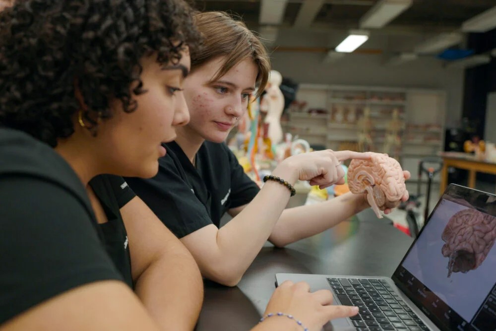
With a swipe of their finger, students can twirl images of a real heart, brain or lungs. They can use voice commands to locate specific anatomical structures or click unfamiliar terms to hear their proper pronunciation. And whether in the classroom or at home, they can add drawings and notes to prepare for upcoming dissections and exams.
“Our goal was to create the best product for our students,” said Alejandro Carrillo, a laboratory instructor and member of the DOCTOR team. “It was inspired by students, and we wanted to create it for students.”
Years in the making and unveiled in late 2024, DOCTOR fills the gap between the static, idealized illustrations of anatomy textbooks and the expensive, proprietary technologies used to teach the subject in schools. It’s still a work in progress, with the team building new features and conducting more dissections and scans.
“The DOCTOR app is different from other software that is out there on the market, because we are using the same resources that we have in the classroom,” said Tomer Kanan, clinical assistant professor of kinesiology and nutrition at UIC and project leader. “It’s not illustration; it’s the real thing. And what students see in the classroom, that’s exactly what they have on the app.”
From the classroom to your device
The inspiration for the app was Kanan’s experience running one of UIC’s largest classes. Each year, more than 700 undergraduates — many planning on careers as doctors, nurses, therapists or other health professionals — take his anatomy and physiology course.
The course is taught in a dry lab, where students learn from intricate plastic models of organs, cells and other structures, and a wet lab, where students do dissection. Photography is not permitted in the wet lab, so students often use their phones to photograph the dry-lab models from multiple angles and create study guides for future exams.
“We found out that students learn best when they own the material in their way, in the way that makes sense to them,” Kanan said. “We started to think, what can we do to take the real world, take what we have in the classroom, and yet give students the ability to control how they want to look at things?”
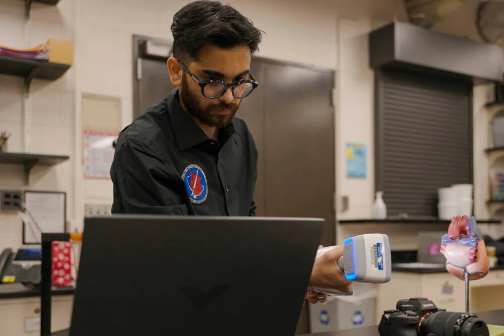
That led Kanan to envision a library of three-dimensional images that students could interact with in an app. He teamed up with Balaji Kashyap Vellaluru, a graduate of the UIC MS in Computer Science program, and they both started learning photogrammetry, the process of scanning and creating 3D images.
Photogrammetry requires hundreds of photographs, methodically taken at precise intervals around an object.
“You have to know exactly what picture you’re taking from which angle, and you always have to keep in mind the three or four previous images that you took,” Vellarulu said. “It was really physically demanding.”
After capturing the images, Vellarulu led the design, development and deployment of the DOCTOR app, implementing advanced interactive features such as custom labeling and voice recognition. More features are on the horizon.
“Whatever you’ve seen so far, it’s nothing compared to what is about to come,” Kanan said.
A puzzle without instructions
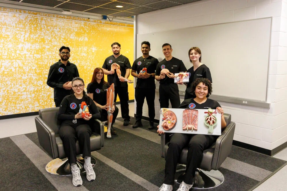
Acquiring the organs to scan is no small task. Fortunately, the DOCTOR team has several skilled dissectors, both practicing health professionals and undergraduates.
UIC students Mirza Baig, Brenda Garcia, Anel Hurtado and Raegan Meyers are former anatomy and physiology students who have stayed involved as dissectors after finishing the course. Each said their experience in Kanan’s class made them want to continue learning anatomy, and they leapt at the opportunity to work on the DOCTOR project team.
“You’re not going to find a group of people that are all so passionate about anatomy, about teaching it and making it accessible (as we are),” Hurtado said. “That’s what I really love about what Dr. Kanan has done here. He has built such an amazing community, it almost feels like a family.”
In addition to designing an apparatus to position cadavers during scans, the students are dissecting the body’s musculature. That work needs to take place between classes, which makes for some unusual text conversations.
“We have a group chat,” Baig said. “We’re like, ‘Hey, do you guys want to hang out and dissect together?’”
For the more complex dissections, Kanan turns to laboratory instructors Carrillo and Victoria Peterson. Carrillo, an occupational therapist, and Peterson, a chiropractor, have clinical experience but also enjoy the teaching and practice of dissection.
“Once you get started, it’s like a really big puzzle,” Peterson said. “There’s not always instructions or a map to follow.”
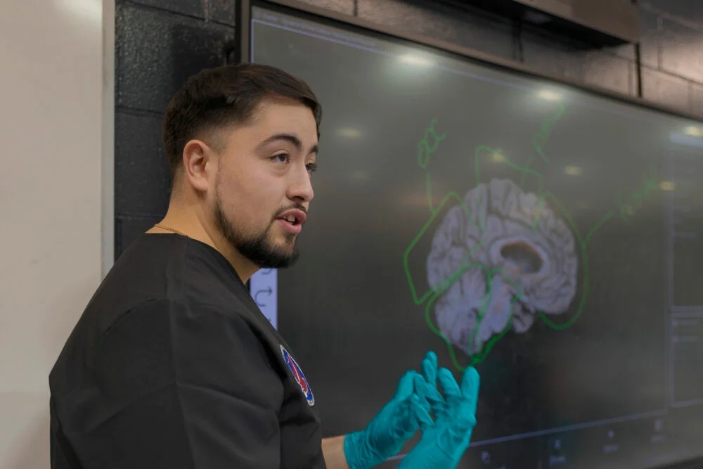
The two tackled Kanan’s biggest challenge so far: a brain with the eyes and spinal cord attached, including the nerves radiating from the organs. The dissection took six months of nights and weekends to complete. But its 3D scan illustrates the ambition of the app, to show complex anatomic structures on an interactive platform.
“I think it’s a great resource, and I really wish I had it when I was in anatomy two years ago,” Baig said. “When I first saw the app, I barged into Dr. Kanan’s office and told him I was happy and jealous at the same time.”
The dead teach the living, again
A decal on a wall of the UIC anatomy and physiology lab spells out a Latin phrase: “Mortui Vivos Docent,” or “The dead teach the living.” That philosophy has motivated human dissection for thousands of years.
But the ancient Greeks didn’t have the technology on the other side of the UIC lab: two big flatscreen TVs. Standing to either side of a TV displaying a 3D image of the human brain, laboratory instructors Carrillo and Peterson rotate the organ and quiz students on its structures. When a student answers, the image zooms in on the corresponding area, allowing for easy comparison between the screen and the real brain on their dissection table.
The DOCTOR app didn’t take long to become an integral part of UIC’s anatomy classes. When the team introduced it to students in the fall 2024 semester, they got a round of applause. Instructors immediately saw students using the app during class. The audio features are especially helpful, they said, as students struggle with the technical, Latin-based terms for many anatomical structures.
After presenting the app at UIC’s Spark Talks in November 2024, Kanan heard from other UIC colleges about using the app in their graduate programs as well. The app is currently available to anyone with a UIC or University of Illinois System account, and he is working with the Office of Technology Management on licensing it to more universities.
“A lot of times, I know that students know the answer I’m looking for when I’m asking questions, but I find some of them are hesitant if they don’t think that they’re going to pronounce it correctly,” Peterson said. “I think it gives students confidence to be able to speak up in class and reinforce their learning that way.”
But Kanan stressed that he will only allow the app to be used by others if they agree to his price for students: free.
“For future clinicians, for people involved with anything regarding the human body, we want to make sure that we give them the best tools to succeed,” Kanan said. “And what better way to do that than to create something that’s never been done, and better yet, to make it free.”